
Optical Imaging Techniques Applied to Dermatology
DOI:
https://doi.org/10.17488/RMIB.45.2.7Keywords:
dermatology, imaging, opticAbstract
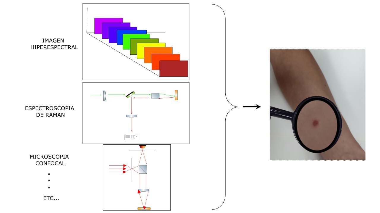
The use of optical imaging technology in recent years has improved diagnostics in the dermatology area. The techniques that can be named are optical coherence tomography, Raman spectroscopy, and optoacoustic imaging. Each technique has applications in general medicine and dermatology but with their respective limitations and focus. This work reviews the imaging techniques in general, prioritizing the theoretical background that allows the technique to operate, and then an image example obtained with the technique is shown. Then, an extensive review of the techniques of hyperspectral imaging and hyperspectral polarized dermatoscope is made. Finally, we talk about new trends in optical imaging future.
Downloads
References
G. Micali, F. Lacarrubba, D. Massimino, R. A. Schwartz, “Dermatoscopy: Alternative uses in daily clinical practice,” J. Am. Acad. Dermatol., vol. 64, no. 6. pp. 1135–1146, 2011, doi: https://doi.org/10.1016/j.jaad.2010.03.010
D. Hirokawa y J. B. Lee, “Dermatoscopy: An overview of subsurface morphology,” Clin. Dermatol., vol. 29, no. 5, pp. 557–565, 2011, doi: https://doi.org/10.1016/j.clindermatol.2010.12.002
C. Benvenuto-Andrade, S. W. Dusza, A. L. Agero, A. Scope, M. Rajadhyaksha, A. C. Halpern, A. A. Marghoob, “Differences Between Polarized Light Dermoscopy and Immersion Contact Dermoscopy for the Evaluation of Skin Lesions,” Arch. Dermatol., vol. 143, no. 3, pp. 329-338, 2007, doi: https://doi.org/10.1001/archderm.143.3.329
V. Tuchyn, Tissue Optics, Bellingham, Estados Unidos: Society of Photo-Optical Instrumentation Engineers, 2015.
R. Splinter, B. A. Hooper, An Introduction to Biomedical Optics, Estados Unidos, CRC Press, 2006.
E. Hecht, Optics, Estados Unidos: Pearson Education, 2017.
D. Malacara, Óptica básica, México: Fondo de Cultura Económica, 2015.
J. Olsen, J. Holmes, G. B. E. Jemec, “Advances in optical coherence tomography in dermatology—a review,” J. Biomed. Opt., vol. 23, no. 04, pp. 1-10, 2018, doi: https://doi.org/10.1117/1.jbo.23.4.040901
J. Welzel, S. Schuh, “Noninvasive diagnosis in dermatology,” J. Dtsch. Dermatol. Ges., vol. 15, no. 10, pp. 999–1016, 2017, doi: https://doi.org/10.1111/ddg.13347
E. Sattler, R. Kästle, y J. Welzel, “Optical coherence tomography in dermatology”, J. Biomed. Opt., vol. 18, no. 6, 2013, art. no. 061224, doi: https://doi.org/10.1117/1.jbo.18.6.061224
X. Shu, L. Beckmann, y H. F. Zhang, “Visible-light optical coherence tomography: a review,” J. Biomed. Opt., vol. 22, no. 12, pp. 1-14, 2017, doi: https://doi.org/10.1117/1.jbo.22.12.121707
N. Chuchvara, B. Rao, y X. Liu, “Manually scanned single fiber optical coherence tomography for skin cancer characterization,” Sci. Rep., vol. 11, no. 1, 2021, art. no. 15570, doi: https://doi.org/10.1038/s41598-021-95118-z
C. E. Psomadakis, N. Marghoob, B. Bleicher, y O. Markowitz, “Optical coherence tomography,” Clin. Dermatol., vol. 39, no. 4, pp. 624–634, 2021, doi: https://doi.org/10.1016/j.clindermatol.2021.03.008
T. Gambichler, A. Pljakic, y L. Schmitz, “Recent advances in clinical application of optical coherence tomography of human skin,” Clin. Cosmet. Investig. Dermatol., vol. 8., pp. 345–354, 2015, doi: https://doi.org/10.2147/CCID.S69119
L. M. C. Vasquez-Pinto, E. P. Maldonado, M. P. Raele, M. M. Amaral, y A. Z. de Freitas, “Optical coherence tomography applied to tests of skin care products in humans - a case study,” Skin Res. Technol., vol. 21, no. 1, pp. 90–93, 2015, doi: https://doi.org/10.1111/srt.12161
A. Taruttis y V. Ntziachristos, “Advances in real-time multispectral optoacoustic imaging and its applications,” Nat. Photon., vol. 9, no. 4, pp. 219–227, 2015, doi: https://doi.org/10.1038/nphoton.2015.29
I. A. Bratchenko, V. P. Sherendak, O. O. Myakinin, D. N. Artemuev, et al., “In vivo hyperspectral imaging of skin malignant and benign tumors in visible spectrum,” J. Biomed. Photonics Eng., vol. 4, no. 1, 2018, art. no. 010301, doi: http://dx.doi.org/10.18287/JBPE17.04.010301
R. Abdlaty, J. Orepoulos, P. Sinclair, R. Berman, y Q. Fang, “High throughput AOTF hyperspectral imager for randomly polarized light,” Photonics, vol. 5, no. 1, 2018, art. no. 3, doi: https://doi.org/10.3390/photonics5010003
Y. Matsumoto, Y, Asao, A. Yoshikawa, H, Sekiguchi, et al., “Label-free photoacoustic imaging of human palmar vessels: A structural morphological analysis,” Sci. Rep., vol. 8, no. 1, 2018, art. no. 786, doi: https://doi.org/10.1038/s41598-018-19161-z
O. A. Melsitov, V. P. Sherendak, S. G. Konovalov, y O. O. Myakinin, “Automatic Malignant Melanoma recognition using a Dermatoscopy Imaging Tool,” presentado en IV Conferencia de Conferencias y Escuelas de Modificaciones «Información sobre tecnologías y nanotecnologías» (ИТНТ-2018), Samara, Rusia, 2018. http://repo.ssau.ru/bitstream/Informacionnye-tehnologii-i-nanotehnologii/Automatic-Malignant-Melanoma-recognition-using-a-Dermatoscopy-Imaging-Tool-69115/1/paper_125.pdf
S. J. Leavesley, M. Walters, C. Lopez, T. Baker, et al., “Hyperspectral imaging fluorescence excitation scanning for colon cancer detection,” J. Biomed. Opt., vol. 21, no. 10, 2016, art. no. 104003, doi: https://doi.org/10.1117/1.jbo.21.10.104003
D. Jiang, S. Christ, D. Correa-Gallegos, P. Ramesh, et al., “Injury triggers fascia fibroblast collective cell migration to drive scar formation through N-cadherin,” Nat. Commun., vol. 11, no. 1, 2020, art. no. 5653, doi: https://doi.org/10.1038/s41467-020-19425-1
C. Petrokilidou, G. Gaitanis, I. D. Bassukas, A. Velegraki, E. Guevara, M. Z. Vardaki, N. Kourkoumelis, “Emerging optical techniques for the diagnosis of onychomycosis,” Appl. Sci., vol. 10, no. 7, 2020, art. no. 2340, doi: https://doi.org/10.3390/app10072340
V. K. Ortner, W. Franco, M. Haedersdal, y P. A. Philipsen, “Noninvasive Assessment of Mycotic Nail Tissue Using an Ultraviolet Fluorescence Excitation Imaging System,” Lasers Surg. Med., vol. 53, no. 2, pp. 245–251, 2021, doi: https://doi.org/10.1002/lsm.23285
A. P. da Silva, T. C. Fortunato, M. D. Stringasci, C. Kurachi, V. S. Bagnato, y N. M. Inada, “Onychomycosis diagnosis using fluorescence and infrared imaging systems”, presentado en Biophotonics South America, Rio de Janeiro, Brasil, 2015, doi: https://doi.org/10.1117/12.2180998
J. Zhao, H. Zeng, S. Kalia, y H. Lui, “Using Raman Spectroscopy to Detect and Diagnose Skin Cancer In Vivo,” Dermatol. Clin., vol. 35, no. 4, pp. 495–504, 2017, doi: https://doi.org/10.1016/j.det.2017.06.010
F. Martelli, S. del Bianco, A. Ismaelli, G. Zaccanti, Light propagation through biological tissue and other diffusive media: theory, solutions, and software. Estados Unidos: SPIE - The International Society for Optical Engineering, 2009.
D. Lunter, V. Klang, D. Kocsis, Z. Varga-Medveczky, S. Berkó, y F. Erdő, “Novel aspects of Raman spectroscopy in skin research,” Exp. Dermatol., vol. 31, no. 9, pp. 1311–1329, 2022, doi: https://doi.org/10.1111/exd.14645
P. Rostron, S. Gaber, D. Gaber, “Raman Spectroscopy, a review,” Int. J. Eng. Technical Res., vol. 6, no. 1, pp. 2454-4698, 2016.
N. Kuhar, S. Sil, T. Verma, y S. Umapathy, “Challenges in application of Raman spectroscopy to biology and materials,” RSC Adv., vol. 8, no. 46, pp. 25888–25908, 2018, doi: https://doi.org/10.1039/c8ra04491k
H. Wang, A. M. D. Lee, H. Lui, D. I. McLean, y H. Zeng, “A Method for accurate in vivo micro-Raman spectroscopic measurements under guidance of advanced microscopy imaging,” Sci. Rep., vol. 3, 2013, art. no. 1890, doi: https://doi.org/10.1038/srep01890
A. Azan, P. J. Caspers, T. C. Bakker Schut, S. Roy, et al., “A novel spectroscopically determined pharmacodynamic biomarker for skin toxicity in cancer patients treated with targeted agents,” Cancer Res., vol. 77, no. 2, pp. 557–565, 2017, doi: https://doi.org/10.1158/0008-5472.can-16-1733
N. Chaudhary, C. Wynne, y A. D. Meade, “A review of applications of Raman spectroscopy in immunology,” Biomed. Spectrosc. Imaging, vol. 9, no. 1–2, pp. 23–31, 2020, doi: https://doi.org/10.3233/BSI-200198
R. Cabrera-Alonso, E. Guevara, M. G. Ramírez-Elías, B. Moncada, y F. J. González, “Detection of hydroquinone by Raman spectroscopy in patients with melasma before and after treatment,” Skin Res. Technol., vol. 25, no. 1, pp. 20–24, 2019, doi: https://doi.org/10.1111/srt.12589
E. Cinotti, J. L. Perrot, B. Labeille, F. Cambazard, y P. Rubegni, “Ex vivo confocal microscopy: an emerging technique in dermatology,” Dermatol. Pract. Concept., vol. 8, no. 2, pp. 109–119, 2018, doi: https://doi.org/10.5826/dpc.0802a08
H. J. Ahn, H. J. Kim, H. Ham, J. H. Baek, et al., “Visualizing the in-vivo application of zinc in sensitive skin using reflectance confocal microscopy,” Sci. Rep., vol. 11, no. 1, 2021, art. no. 7738, doi: https://doi.org/10.1038/s41598-021-87346-0
E. Guevara, J. Manuel Gutierrez-Hernandez, A. Castonguay, F. Lesage, B. Moncada, y F. J. González, “Morphological and molecular imaging of skin simples,” Biomed. Res., vol. 28, núm. 4, 2017. [En línea]. Disponible en: https://www.alliedacademies.org/articles/morphological-and-molecular-imaging-of-skin-samples.html
D. H. Goldstein, Polarized light, 3ra ed. Estados Unidos: CRC press, 2017.
C. Whybrew, P. Pietkiewicz, I. Kohut, J. C. Chia, B. N. Akay, y C. Rosendahl, “Not All Polarized-light Dermatoscopes May Display Diagnostically Critical Polarizing-specific Features,” Dermatol. Pract. Concept., vol. 12, no. 4, 2022, art. no. e2022250, doi: https://doi.org/10.5826/dpc.1204a250
A. Nkengne, J. Robic, P. Seroul, S. Gueheunneux, M. Jomier, y K. Vie, “SpectraCam®: A new polarized hyperspectral imaging system for repeatable and reproducible in vivo skin quantification of melanin, total hemoglobin, and oxygen saturation,” Skin Res. Technol., vol. 24, núm. 1, pp. 99–107, 2018, doi: https://doi.org/10.1111/srt.12396
F. Vasefi, N. MacKinnon, R. Saager, K. M. Kelly, et al., “Multimode optical dermoscopy (SkinSpect) analysis for skin with melanocytic nevus,” presentado en Imaging, Manipulation, and Analysis of Biomolecules, Cells, and Tissues IX, SPIE, San Francisco, California, Estados Unidos, 2016, art. no. 971110. doi: https://doi.org/10.1117/12.2214288
M. J. Khan, H. S. Khan, A. Yousaf, K. Khurshid, y A. Abbas, “Modern Trends in Hyperspectral Image Analysis: A Review,” IEEE Access, vol. 6, pp. 14118–14129, 2018, doi: https://doi.org/10.1109/ACCESS.2018.2812999
J. Yoon, J. Joseph, D. J. Waterhouse, A. S. Luthman, et al., “A clinically translatable hyperspectral endoscopy (HySE) system for imaging the gastrointestinal tract,” Nat. Commun., vol. 10, no. 1, 2019, art. no. 1902, doi: https://doi.org/10.1038/s41467-019-09484-4
A. M. Hosking, B. J. Coakley, D. Chang, F. Talebi-Liasi, et al., “Hyperspectral imaging in automated digital dermoscopy screening for melanoma,” Lasers Surg. Med., vol. 51, no. 3, pp. 214–222, 2019, doi: https://doi.org/10.1002/lsm.23055
L. Rey-Barroso, F. J. Burgos-Fernández, X. Delpueyo, M. Ares, et al., “Visible and extended near-infrared multispectral imaging for skin cancer diagnosis,” Sensors, vol. 18, no. 5, 2018, art. no. 1441, doi: https://doi.org/10.3390/s18051441
N. Neittaanmäki-Perttu, M. Grönroos, L. Jeskanen, I. Pölönen, A. Ranki, O. Saksela, E. Snellman, “Delineating margins of lentigo maligna using a hyperspectral imaging system,” Acta Derm. Venereol., vol. 95, no. 5, pp. 549–552, 2015, doi: https://doi.org/10.2340/00015555-2010
H. Fabelo, V. Melián, B. Martínez, P. Beltrán, et al., “Dermatologic Hyperspectral Imaging System for Skin Cancer Diagnosis Assistance,” presentado en 2019 XXXIV Conference on Design of Circuits and Integrated Systems (DCIS), Bilbao, España, 2019, pp. 1–6, doi: https://doi.org/10.1109/DCIS201949030.2019.8959869
Y. Gu, Y.-P. Partridge, y J. Zhou, “A Hyperspectral Dermoscopy Dataset for Melanoma Detection,” presentado en OR 2.0 Context-Aware Operating Theaters, Computer Assisted Robotic Endoscopy, Clinical Image-Based Procedures, and Skin Image Analysis, Granada, España, 2018, pp. 268–276, doi: https://doi.org/10.1007/978-3-030-01201-4_29
E. J. M. Baltussen, E. N. D. Kok, S. G. Brouwer de Koning, J. Sanders, et al., “Hyperspectral imaging for tissue classification, a way toward smart laparoscopic colorectal surgery,” J. Biomed. Opt., vol. 24, no. 1, pp. 1-9, 2019, doi: https://doi.org/10.1117/1.jbo.24.1.016002
S. Ortega, M. Halicek, H. Fabelo, R. Guerra, et al., “Hyperspectral imaging and deep learning for the detection of breast cancer cells in digitized histological images,” Proc. SPIE Int. Soc. Opt. Eng., 2020, art. no. 113200V, doi: https://doi.org/10.1117/12.2548609
E. Zherebtsov, A. Popov, A. Doronin, I. Meglinski, y A. Bykov, “Hyperspectral system for imaging of skin chromophores and blood oxygenation,” presentado en Diffuse Optical Spectroscopy and Imaging VI 2017, Munich, Alemania, 2017, doi: https://doi.org/10.1117/12.2280779
Q. He y R. Wang, “Hyperspectral imaging enabled by an unmodified smartphone for analyzing skin morphological features and monitoring hemodynamics,” Biomed. Opt. Express, vol. 11, no. 2, pp. 895-910, 2020, doi: https://doi.org/10.1364/boe.378470
A. Kulcke, A. Holmer, P. Wahl, F. Siemers, T. Wild, y G. Daeschlein, “A compact hyperspectral camera for measurement of perfusion parameters in medicin,e” Biomed. Tech., vol. 63, no. 5, pp. 547–556, 2018, doi: https://doi.org/10.1515/bmt-2017-0145
E. Zherebtsov, V. Dremin, A. Popov, A. Doronin, et al., “Hyperspectral imaging of human skin aided by artificial neural networks,” Biomed. Opt. Express, vol. 10, no. 7, pp. 3545-3559, 2019, doi: https://doi.org/10.1364/boe.10.003545
O. Abeyakoon, R. Woitek, M. G. Wallis, P. L. Moyle, et al., “An optoacoustic imaging feature set to characterise blood vessels surrounding benign and malignant breast lesions,” Photoacoustics, vol. 27, 2022, art. no. 100383, doi: https://doi.org/10.1016/j.pacs.2022.100383
A. F. Kukk, F. Scheling, R. Panzer, S. Emmert, y B. Roth, “Combined ultrasound and photoacoustic C-mode imaging system for skin lesion assessment,” Sci. Rep., vol. 13, no. 1, 2023, art. no. 17947, doi: https://doi.org/10.1038/s41598-023-44919-5
J. Kukačka, S. Metz, C. Dehner, A. Muckenhuber, et al., “Image processing improvements afford second-generation handheld optoacoustic imaging of breast cancer patients,” Photoacoustics, vol. 26, 2022, art. no. 100343 doi: https://doi.org/10.1016/j.pacs.2022.100343
T. Nau, C. Schönmann, B. Hindelang, L. Riobo, et al., “Raster-scanning optoacoustic mesoscopy biomarkers for atopic dermatitis skin lesions,” Photoacoustics, vol. 31, 2023, art. no. 100513, doi: https://doi.org/10.1016/j.pacs.2023.100513
L. Yan, S. Hu, A. Alzahrani, S. Alharbi, y P. Blanos, “A multi-wavelength opto-electronic patch sensor to effectively detect physiological changes against human skin types,” Biosensors, vol. 7, no. 2, 2017, art. no. 22, doi: https://doi.org/10.3390/bios7020022
F. Tanriverdi, D. Schuldt, y J. Thiem, “Hyperspectral Imaging: Color Reconstruction Based on Medical Data,” presentado en 2018 IEEE-EMBS Conference on Biomedical Engineering and Sciences (IECBES), Sarawak, Malaysia, 2018, pp. 194–199, doi: https://doi.org/10.1109/IECBES.2018.8626614
D. Salo, H. Zhang, D. M. Kim, M. Y. Berezin, “Multispectral measurement of contrast in tissue-mimicking phantoms in near-infrared spectral range of 650 to 1600 nm,” J. Biomed. Opt., vol. 19, no.8, 2014, art. no. 086008, doi: https://doi.org/10.1117/1.jbo.19.8.086008
A. Jullien, R. Pascal, U. Bortolozzo, N. Forget, y S. Residori, “High-resolution hyperspectral imaging with cascaded liquid crystal cells,” Optica, vol. 4, no. 4, pp. 400-405, 2017, doi: https://doi.org/10.1364/OPTICA.4.000400
J. Pichette, W. Charle, y A. Lambrechts, “Fast and compact internal scanning CMOS-based hyperspectral camera: the Snapscan,” presentado en Photonic Instrumentation Engineering IV, San Francisco California, Estados Unidos, 2017, art. no. 1011014, doi: https://doi.org/10.1117/12.2253614
K. B. Yushkov y V. Ya. Molchanov, “Hyperspectral imaging acousto-optic system with spatial filtering for optical phase visualization,” J. Biomed. Opt., vol. 22, no. 6, 2017, art. no. 066017, 2017, doi: https://doi.org/10.1117/1.jbo.22.6.066017
B. Nirmal, A. Krishnaram, y R. Sudhagar, “Rainbow sign in dermatoscopy of nodular basal cell carcinoma,” Indian J. Dermatopathol. Diagn. Dermatol., vol. 6, no. 2, pp. 107-108, 2019, doi: https://doi.org/10.4103/ijdpdd.ijdpdd_27_19
D. Kapsokalyvas, R. Cicchi, N. Bruscino, D. Alfieri, et al., “In-vivo imaging of psoriatic lesions with polarization multispectral dermoscopy and multiphoton microscopy,” Biomed. Opt. Express, vol. 5, no. 7, pp. 2405-2419, 2014, doi: https://doi.org/10.1364/boe.5.002405
T. Van Tien, N. Hoang Ohuc, L. Quang Nhien, T. Thi Thu Trang, et al., “Evaluation of scaly levels in psoriasis using multispectral polarized imaging,” presentado en 6th International Conference on the Development of Biomedical Engineering in Vietnam (BME6), Vietnam, 2018, pp. 97–101, doi: https://doi.org/10.1007/978-981-10-4361-1_16
S. G. Konovalov, O. A. Melsitov, O. O. Myakinin, I. A. Bratchenko, A. A. Moryatov, S. V. Kozlov, V. P Zakharov, “Dermatoscopy software tool for in vivo automatic malignant lesions detection,” J. Biomed. Photonics, Eng., vol. 4, no. 4, pp. 105–118, 2018, doi: http://dx.doi.org/10.18287/JBPE18.04.040302
D. Kapsokalyvas, N. Bruscino, D. Alfieri, V. de Giorgi, et al., “Spectral morphological analysis of skin lesions with a polarization multispectral dermoscope,” Opt. Express, vol. 21, no. 4, pp. 4826–4840, 2013, doi: https://doi.org/10.1364/oe.21.004826
Q. He y R. K. Wang, “Analysis of skin morphological features and real-time monitoring using snapshot hyperspectral imaging,” Biomed. Opt. Express, vol. 10, no. 11, pp. 5625-5638, 2019, doi: https://doi.org/10.1364/boe.10.005625
V. Dremin, A. Bykov, Z. Marcinkevics, A. Grabovskis, E. Zherebtsov, A. Popov, I. Meglinski, “Assessment of Age-related Skin Changes Using Hyperspectral Polarization Imaging,” presentado en Medical Laser Applications and Laser-Tissue Interactions IX, Munich, Alemania, 2019, art. no. 110718, doi: https://doi.org/10.1117/12.2526359
D. Fricke, M. Wollweber, y B. Roth, “Mueller Matrix Measurement System for Skin Polarimetry as Additional Module for Non-Contact Dermatoscopy,” presentado en 2019 Conference on Lasers and Electro-Optics Europe & European Quantum Electronics Conference (CLEO/Europe-EQEC), Muchich, Alemania, 2019, pp. 1-1, doi: https://doi.org/10.1109/CLEOE-EQEC.2019.8872705
Q. Wang, J. Shi, J. Wang, D. Zhao, y Y. Liu, “Design and Characterization of an AOTF Hyper-Spectral Polarization Imaging System,” J. Mod. Opt., vol. 64, no. 1, pp. 1–7, 2017, doi: https://doi.org/10.1080/09500340.2016.1200682
J. L. Xu, A. Gobrecht, N. Gorretta, D. Héran, A. A. Gowen, y R. Bendoula, “Development of a polarized hyperspectral imaging system for investigation of absorption and scattering properties,” J. Near Infrared Spectrosc., vol. 27, no. 4, pp. 314–329, 2019, doi: https://doi.org/10.1177/0967033519857732
J. Olsen, P. Lindsø Andersen, L. Themstrup, G. B. E. Jemec, y D. M. L. Saunte, “Optical coherence tomography of onychomycosis: proposed terminology and a suggestion of practical usage,” Arch. Dermatol. Res., vol. 312, no. 1, pp. 51–58, 2020, doi: https://doi.org/10.1007/s00403-019-01989-8
I. Steinberg, D. M. Huland, O. Vermesh, H. E. Frostig, W. S. Tummers, y S. S. Gambhir, “Photoacoustic clinical imaging,” Photoacoustics, vol. 14, pp. 77–98, 2019, doi: https://doi.org/10.1016/j.pacs.2019.05.001
D. R. Miller, J. W. Jarrett, A. M. Hassan, y A. K. Dunn, “Deep tissue imaging with multiphoton fluorescence microscopy,” Curr. Opin. Biomed. Eng., vol. 4, pp. 32–39, 2017, doi: https://doi.org/10.1016/j.cobme.2017.09.004
S. L. P. Aggarwal y F. A. Papay, “Applications of multispectral and hyperspectral imaging in dermatology,” Exp. Dermatol., vol. 31, no. 8, pp. 1128–1135, 2022, doi: https://doi.org/10.1111/exd.14624
J. Yoon, “Hyperspectral Imaging for Clinical Applications,” Biochip J., vol. 16, no. 1, 2022, doi: https://doi.org/10.1007/s13206-021-00041-0
A. Orlando, F. Frranceschini, C. Muscas, S. Pidkova, M. Bartolini, M. Rovere, A. Tagliaferro, “A comprehensive review on Raman spectroscopy applications,” Chemosensors, vol. 9, no. 9, 2021, art. no. 262, doi: https://doi.org/10.3390/chemosensors9090262
S. Duraipandian, M. Sylvest Bergholt, W. Zheng, K. Yu Ho, M. Teh, K. Guan Yeoh, J. Bok Yan So, A. Shabbir, Z. Huang, “Real-time Raman spectroscopy for in vivo, online gastric cancer diagnosis during clinical endoscopic examination,” J. Biomed. Opt., no. 8, 2012, art. no. 081418, doi: https://doi.org/10.1117/1.jbo.17.8.081418
A. D. Elliott, “Confocal Microscopy: Principles and Modern Practices,” Curr. Protoc. Cytom., vol. 92, no. 1, 2020, e68, doi: https://doi.org/10.1002/cpcy.68
S. Y. Chen, Z.-T. Su, D.-J. Lin, M.-X. Lee, et al., “Optimizing imaging depth of anisotropic scattering tissues with polarization engineered second harmonic generation microscopy,” Results Phys., vol. 28, 2021, art. no. 104653, doi: https://doi.org/10.1016/j.rinp.2021.104653
Downloads
Published
How to Cite
Issue
Section
License
Copyright (c) 2024 Revista Mexicana de Ingenieria Biomedica

This work is licensed under a Creative Commons Attribution-NonCommercial 4.0 International License.
Upon acceptance of an article in the RMIB, corresponding authors will be asked to fulfill and sign the copyright and the journal publishing agreement, which will allow the RMIB authorization to publish this document in any media without limitations and without any cost. Authors may reuse parts of the paper in other documents and reproduce part or all of it for their personal use as long as a bibliographic reference is made to the RMIB. However written permission of the Publisher is required for resale or distribution outside the corresponding author institution and for all other derivative works, including compilations and translations.


