
A U-Net with Statistical Shape Restrictions Applied to the Segmentation of the Left Ventricle in Echocardiographic Images
DOI:
https://doi.org/10.17488/RMIB.44.4.10Keywords:
convolutional neural networks, echocardiography, Left ventricle segmentation, statistical shape analysisAbstract
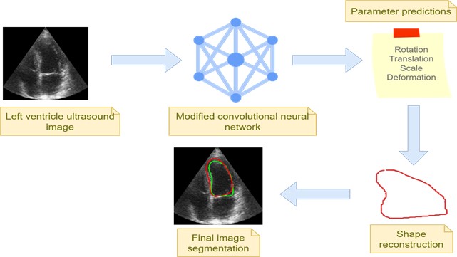
This paper aims to introduce an innovative approach to semantic segmentation by leveraging a convolutional neural network (CNN) for predicting the shape and pose parameters of the left ventricle (LV). Our approach involves a modified U-Net architecture with a regression layer as the final stage, as opposed to the traditional classification layer. This modification allows us to predict all the shape and pose parameters of a statistical shape model, including rotation, translation, scale, and deformation. The adapted U-Net is trained using data from a point distribution model (PDM) of the LV. The experimental results demonstrate a mean Dice coefficient of 0.82 on good quality images, and 0.66 including mean and low-quality images. Our approach successfully overcomes a common issue encountered in CNN-based semantic segmentation. Unlike the inaccurate pixel classification that often leads to unwanted blobs, our CNN generates statistically valid shapes. These shapes hold significant potential in initializing other methods, such as active shape models (ASMs). Our novel CNN-based approach provides a novel solution for semantic segmentation, offering shapes and pose parameters that can enhance the accuracy and reliability of subsequent medical image analysis methods.
Downloads
References
N. Paragios and R. Deriche, “Geodesic active regions and level set methods for supervised texture segmentation,” Int. J. Comput. Vis., vol. 46, no. 3, pp. 223–247, Feb. 2002, doi: https://doi.org/10.1023/A:1014080923068
N. Paragios, “A level set approach for shape-driven segmentation and tracking of the left ventricle,” IEEE Trans. Med. Imag., vol. 22, no. 6, pp. 773–776, Jun. 2003, doi: https://doi.org/10.1109/TMI.2003.814785
J. C. Nascimento and J. S. Marques, “Robust shape tracking with multiple models in ultrasound images,” IEEE Trans. Image Process., vol. 17, no. 3, pp. 392–406, Mar. 2008, doi: https://doi.org/10.1109/TIP.2007.915552
V. Zagrodsky, V. Walimbe, C. R. Castro-Pareja, J. X. Qin, J.-M. Song, and R. Shekhar, “Registration-assisted segmentation of real-time 3-D echocardiographic data using deformable models,” IEEE Trans. Med. Imaging., vol. 24, no. 9, pp. 1089–1099, Sep. 2005, doi: https://doi.org/10.1109/tmi.2005.852057
B. Georgescu, X. S. Zhou, D. Comaniciu, and A. Gupta, “Database-guided segmentation of anatomical structures with complex appearance,” in 2005 IEEE Computer Society Conference on Computer Vision and Pattern Recognition (CVPR'05), San Diego, CA, USA, 2005, pp. 429-436 vol. 2, doi: https://doi.org/10.1109/CVPR.2005.119
S. C. Mitchell, B. P. F. Lelieveldt, R. J. van der Geest, H. G. Bosch, J. H. C. Reiber, and M. Sonka, “Multistage hybrid active appearance model matching: Segmentation of left and right ventricles in cardiac MR images,” IEEE Trans. Med. Imag., vol. 20, no. 5, pp. 415–423, May 2001, doi: https://doi.org/10.1109/42.925294
O. Bernard, J. G. Bosch, B. Heyde, M. Alessandrini, et al., “Standardized Evaluation System for Left Ventricular Segmentation Algorithms in 3D Echocardiography,” IEEE Trans. Med. Imaging, vol. 35, no. 4, pp. 967-977, Apr. 2016, doi: https://doi.org/10.1109/TMI.2015.2503890
S. Leclerc, T. Grenier, F. Espinosa and O. Bernard, “A fully automatic and multi-structural segmentation of the left ventricle and the myocardium on highly heterogeneous 2D echocardiographic data,” 2017 IEEE International Ultrasonics Symposium (IUS), Washington, DC, USA, 2017, pp. 1-4, doi: https://doi.org/10.1109/ULTSYM.2017.8092797
T. F. Cootes, C. J. Taylor, D. H. Cooper, J. Graham, “Active shape models their training and application,” Comput. Vis. Image Underst., vol. 61, no. 1, pp. 38–59, Jan. 1995, doi: https://doi.org/10.1006/cviu.1995.1004
Y. Ali, S. Beheshti, F. Janabi-Sharifi, “Echocardiogram segmentation using active shape model and mean squared eigenvalue error,” Biomed. Signal Process. Control, vol. 69, art. no. 102807, Aug. 2021, doi: https://doi.org/10.1016/j.bspc.2021.102807
M. A. Shoaib, J. Huang Chuah, R. Ali, K. Hasikin, et al., “An Overview of Deep Learning Methods for Left Ventricle Segmentation,” Comput. Intell. Neurosci., vol. 2023, art. no. 4208231, 2023, doi: https://doi.org/10.1155/2023/4208231
N. Savioli, M. S. Vieira, P. Lamata, G. Montana, “Automated Segmentation on the Entire Cardiac Cycle Using a Deep Learning Work – Flow,” in 2018 Fifth International Conference on Social Networks Analysis, Management and Security (SNAMS), Valencia, Spain, 2018, pp. 153-158, doi: https://doi.org/10.1109/SNAMS.2018.8554962
X. Zou, Q. Wang, T. Luo, “A novel approach for left ventricle segmentation in tagged MRI,” Comput. Electr. Eng., vol. 95, art. no. 107416, Oct. 2021, doi: https://doi.org/10.1016/j.compeleceng.2021.107416
T. Wech, M. J. Ankenbrand, T. A. Bley, J. F. Heidenreich, “A data-driven semantic segmentation model for direct cardiac functional analysis based on undersampled radial MR cine series,” Magn. Reason. Med., vol. 87, no. 2, pp. 972-983, Feb. 2022, doi: https://doi.org/10.1002/mrm.29017
G. Veni, M. Moradi, H. Bulu, G. Narayan, and T. Syeda-Mahmood, “Echocardiography segmentation based on a shape-guided deformable model driven by a fully convolutional network prior,” 2018 IEEE 15th International Symposium on Biomedical Imaging (ISBI 2018), Washington, DC, USA, 2018, pp. 898-902, doi: https://doi.org/10.1109/ISBI.2018.8363716
W.-Y. Hsu, “Automatic Left Ventricle Recognition, Segmentation and Tracking in Cardiac Ultrasound Image Sequences,” IEEE Access, vol. 7, pp. 140524-140533, 2019, doi: https://doi.org/10.1109/ACCESS.2019.2920957
Y. Li, W. Lu, P. Monkam, Z. Zhu, W. Wu, M. Liu, “LVSnake: Accurate and robust left ventricle contour localization for myocardial infarction detection,” Biomed. Signal Process. Control, vol. 85, art. no. 105076, Aug. 2023, doi: https://doi.org/10.1016/j.bspc.2023.105076
S. Peng, W. Jiang, H. Pi, X. Li, H. Bao, and X. Zhou, “Deep Snake for Real-Time Instance Segmentation,” 2020, arXiv: 2001.01629, doi:
https://doi.org/10.48550/arXiv.2001.01629
O. Ronneberger, P. Fischer, T. Brox, “U-Net: Convolutional Networks for Biomedical Image Segmentation,” Medical Image Computing and Computer-Assisted Intervention – MICCAI 2015, Munich, Germany, 2015, pp. 234-241, doi: https://doi.org/10.1007/978-3-319-24574-4_28
S. Leclerc, E. Smistad, J. Pedrosa, A. Østvik, et al., “Deep Learning for Segmentation using an Open Large-Scale Dataset in 2D Echocardiography,” IEEE Trans. Med. Imaging, vol. 38, no. 9, pp. 2198-2210, Sep. 2019, doi: https://doi.org/10.1109/TMI.2019.2900516
EchoNet-Dynamic: a Large New Cardiac Motion Video Data Resource for Medical Machine Learning, github, 2019. [Online]. Available: https://echonet.github.io/dynamic/index.html#motivation
E. Galicia Gomez, F. Torres Robles, B. Escalante Rmirez, J. Olveres, F. Arámbula Cosío, “Full multi resolution active shape model for left ventricle segmentation,” in 17th International Symposium on Medical Information Processing and Analysis, Campinas, Brazil, 2021, doi: https://doi.org/10.1117/12.2606252
M. B. Stegmann, D. Delgado Gomez. (2002). A Brief Introduction to Statistical Shape Analysis [Online]. Available: http://www2.imm.dtu.dk/pubdb/edoc/imm403.pdf
A. Cervantes-Guzmán, K. McPherson, J. Olveres, C. F. Moreno-García, F. Torres Robles, E. Elyan, B. Escalante-Ramírez, “Robust cardiac segmentation corrected with heuristics,” PLoS One, vol. 18, no. 10, art. no. E0293560, Oct. 2023, doi: https://doi.org/10.1371/journal.pone.0293560
Published
How to Cite
Issue
Section
License
Copyright (c) 2023 Revista Mexicana de Ingenieria Biomedica

This work is licensed under a Creative Commons Attribution-NonCommercial 4.0 International License.
Upon acceptance of an article in the RMIB, corresponding authors will be asked to fulfill and sign the copyright and the journal publishing agreement, which will allow the RMIB authorization to publish this document in any media without limitations and without any cost. Authors may reuse parts of the paper in other documents and reproduce part or all of it for their personal use as long as a bibliographic reference is made to the RMIB. However written permission of the Publisher is required for resale or distribution outside the corresponding author institution and for all other derivative works, including compilations and translations.


