
Optimizing Electrode Performance in EMG and EIT for Superior Muscle Data Acquisition
DOI:
https://doi.org/10.17488/RMIB.46.SI-TAIH.1514Keywords:
EMG, EIT, electrodes, isometric contraction, isotonic contractionAbstract
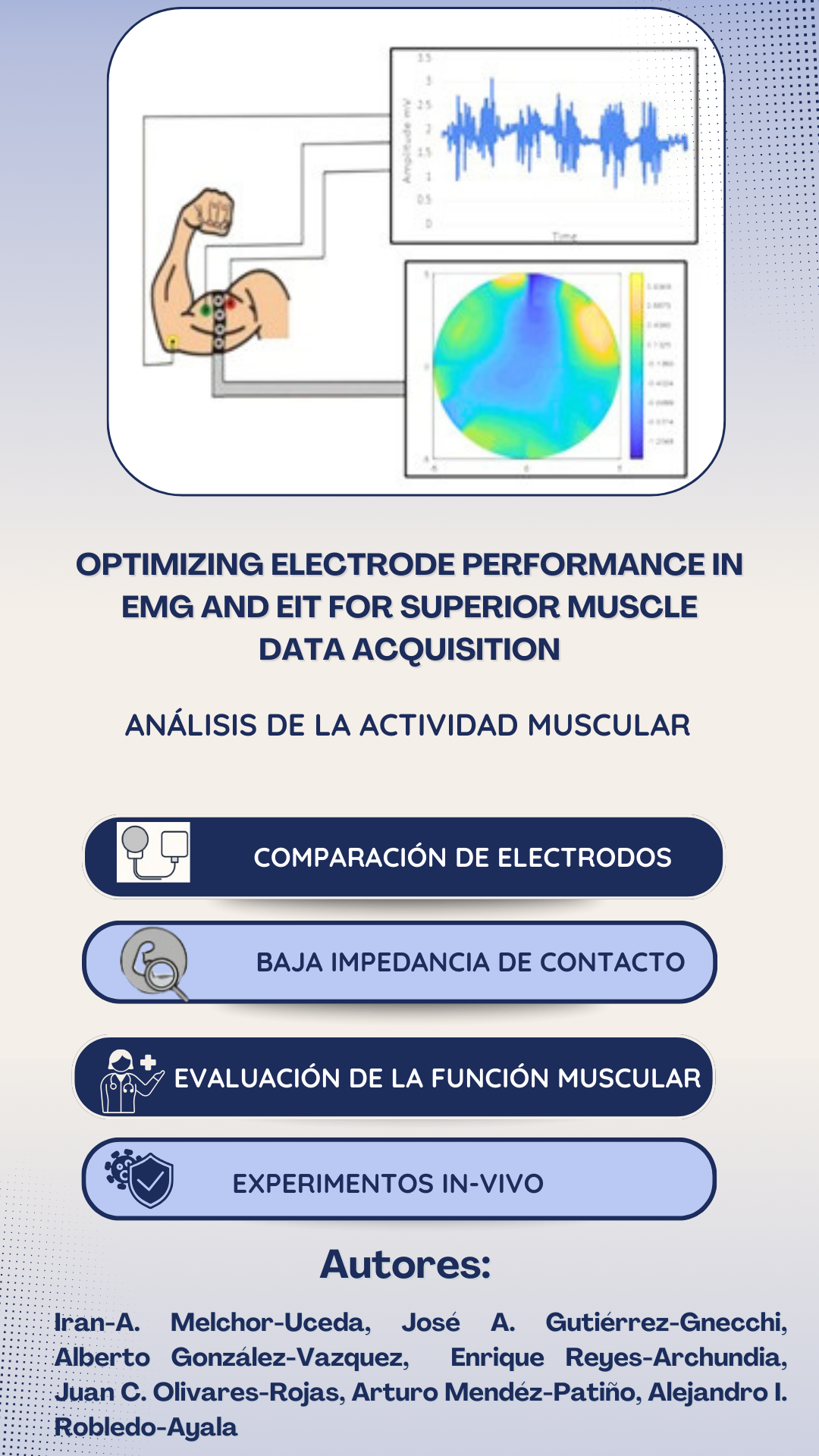
Optimizing electrode performance in electromyography (EMG) and electrical impedance tomography (EIT) is critical to advancing muscle data acquisition. This study systematically evaluates various electrode types, shapes, and materials, focusing on optimizing signal-to-noise ratio, durability, and long-term usability. A key contribution of this research is the identification of stainless-steel electrodes as the most efficient option, demonstrating superior signal stability, oxidation resistance, and reusability compared to disposable alternatives. This finding not only improves the reliability of EMG and EIT measurements but also offers a sustainable and cost-effective solution for clinical and research applications. By providing empirical evidence on electrode selection and design, this study lays the foundation for improved methodologies in rehabilitation, sports medicine, and neurology, ultimately improving patient care and deepening understanding of muscle physiology.
Downloads
References
E. Criswell, Cram’s Introduction to Surface Electromyography, 2nd ed., London, U.K: Jones & Bartlett Learning, 2010.
R. Merletti and P. J. Parker, Electromyography: Physiology, Engineering, and Non-Invasive Applications, 1st ed., Hoboken, New Jersey, U.S.: John Wiley & Sons, 2005.
E. A. Clancy and N. Hogan, “Probability density of the surface electromyogram and its relation to amplitude detectors,” IEEE Trans. Biomed. Eng., vol. 46, no. 6, pp. 730–739, 1999, doi: https://doi.org/10.1109/10.764949
D. Yang, et al., “Dynamic training protocol improves the robustness of PR-based myoelectric control,” Biomed. Signal Process. Control, vol. 31, pp. 249–256, 2017, doi: https://doi.org/10.1016/j.bspc.2016.08.017
M. V. Arteaga, et al., “EMG-driven hand model based on the classification of individual finger movements,” Biomed. Signal Process. Control, vol. 58, 2020, art. no. 101834, doi: https://doi.org/10.1016/j.bspc.2019.101834
L. Bi, A. -->Genetu Feleke and C. Guan, “A review on EMG-based motor intention prediction of continuous human upper limb motion for human-robot collaboration,” Biomed. Signal Process.Control, vol. 51, pp. 113–127, 2019, doi: https://doi.org/10.1016/j.bspc.2019.02.011
R. Wen, et al., “Force-Guided High-Precision Grasping Control of Fragile and Deformable Objects Using sEMG-Based Force Prediction,” IEEE Robot. Automat. Lett., vol. 5, no. 2, pp. 2762–2769, 2020, doi: https://doi.org/10.1109/lra.2020.2974439
A. J. Young, L. J. Hargrove and T. A. Kuiken, “The Effects of Electrode Size and Orientation on the Sensitivity of Myoelectric Pattern Recognition Systems to Electrode Shift,” IEEE Trans. Biomed. Eng., vol. 58, no. 9, pp. 2537–2544, 2011, doi: https://doi.org/10.1109/tbme.2011.2159216
L. Hargrove, K. Englehart and B. Hudgins, “A training strategy to reduce classification degradation due to electrode displacements in pattern recognition based myoelectric control,” Biomed. Signal Process. Control, vol. 3, no. 2, pp. 175–180, 2008, doi: https://doi.org/10.1016/j.bspc.2007.11.005
Y. Geng, P. Zhou and G. Li, “Toward attenuating the impact of arm positions on electromyography pattern-recognition based motion classification in transradial amputees,” J. NeuroEng. Rehabil., vol. 9, no. 1, 2012, art. no. 74, doi: https://doi.org/10.1186/1743-0003-9-74
A. Fougner, et al., “Resolving the Limb Position Effect in Myoelectric Pattern Recognition,” IEEE Trans. Neural Syst. Rehabil. Eng., vol. 19, no. 6, pp. 644–651, 2011, doi: https://doi.org/10.1109/tnsre.2011.2163529
D. Tkach, H. Huang and T. A. Kuiken, “Study of stability of time-domain features for electromyographic pattern recognition”, J. NeuroEng. Rehabil., vol. 7, 2010, art. no. 21, doi: https://doi.org/10.1186/1743-0003-7-21
J. W. Sensinger, B. A. Lock and T. A. Kuiken, “Adaptive Pattern Recognition of Myoelectric Signals: Exploration of Conceptual Framework and Practical Algorithms”, IEEE Trans. Neural Syst. Rehabil. Eng., vol. 17, no. 3, pp. 270–278, 2009, doi: https://doi.org/10.1109/tnsre.2009.2023282
Q. Xi, et al., “A novel localized collocation solver based on Trefftz basis for potential-based inverse electromyography”, Appl. Math. Computation, vol. 390, 2021, art. no. 125604, doi: https://doi.org/10.1016/j.amc.2020.125604
K. van den Doel, U. M. Ascher and D. K. Pai, “Source localization in electromyography using the inverse potential problem”, Inverse Probl., vol. 27, no. 2, 2011, art. no. 025008, doi: https://doi.org/10.1088/0266-5611/27/2/025008
P. S. Cuculich, et al., “Noninvasive real-time mapping of an incomplete pulmonary vein isolation using electrocardiographic imaging”, Heart Rhythm, vol. 7, no. 9, pp. 1316–1317, 2010, doi: https://doi.org/10.1016/j.hrthm.2009.11.009
P. S. Cuculich et al., “The Electrophysiological Cardiac Ventricular Substrate in Patients After Myocardial Infarction,” J. Amer. College Cardiol., vol. 58, no. 18, pp. 1893–1902, 2011, doi: https://doi.org/10.1016/j.jacc.2011.07.029
J. Sun, et al., “Application of Surface Electromyography in Exercise Fatigue: A Review,” Frontiers Syst. Neurosci., vol. 16, Agu. 2022, Art. no. 893275, doi: https://doi.org/10.3389/fnsys.2022.893275
A. J. Young, L. J. Hargrove and T. A. Kuiken, “The Effects of Electrode Size and Orientation on the Sensitivity of Myoelectric Pattern Recognition Systems to Electrode Shift,”,IEEE Trans. Biomed. Eng., vol. 58, no. 9, pp. 2537–2544, 2011, doi: https://doi.org/10.1109/tbme.2011.2159216
I. Morgado, Materia gris: La apasionante historia del conocimiento del cerebro, Spain: Ariel, 2021.
M. Garzón Barrios, et al., “El efecto volta. Un caso de estudio sobre la producción de efectos sensibles y los procesos de teorización en ciencias,” Ens. Pesqui. Em Educ. Em Cienc. (Belo Horizonte), vol. 22, 2020, art. no. e14862, doi: https://doi.org/10.1590/1983-21172020210113
L. K. Huang, et al., “Electrical Impedance Myography Applied to Monitoring of Muscle Fatigue During Dynamic Contractions,” IEEE Access, vol. 8, pp. 13056–13065, 2020, doi: https://doi.org/10.1109/access.2020.2965982
H. F. Hassan, S. J. Abou-Loukh and I. K. Ibraheem, “Teleoperated robotic arm movement using electromyography signal with wearable Myo armband,” J. King Saud Univ. - Eng. Sci., vol. 32, no. 6, pp. 378–387, 2020, doi: https://doi.org/10.1016/j.jksues.2019.05.001
J. N. Wang et al., “Optimization of the electrode configuration of electrical impedance myography for wearable application,” Automatika, vol. 61, no. 3, pp. 475–481, 2020, doi: https://doi.org/10.1080/00051144.2020.1783615
T. J. Freeborn, G. Regard and B. Fu, “Localized Bicep Tissue Bioimpedance Alterations Following Eccentric Exercise in Healthy Young Adults,” IEEE Access, vol. 8, pp. 23100–23109, 2020, doi: https://doi.org/10.1109/access.2020.2970314
T. Freeborn and B. Fu, “Fatigue-Induced Cole Electrical Impedance Model Changes of Biceps Tissue Bioimpedance,” Fractal Fract., vol. 2, no. 4, 2018, art. no. 27, doi: https://doi.org/10.3390/fractalfract2040027
S. B. Rutkove and B. Sanchez, “Electrical Impedance Methods in Neuromuscular Assessment: An Overview,” Cold Spring Harb. Perspect. Med., vol. 9, no. 10, 2018, art. no. a034405, doi: https://doi.org/10.1101/cshperspect.a034405
R. Merletti, et al., “Advances in Surface EMG: Recent Progress in Detection and Processing Techniques,” Crit. Rev. Biomed. Eng., vol. 38, no. 4, pp. 305–345, 2010, doi: https://doi.org/10.1615/critrevbiomedeng.v38.i4.10
M. Rinaldi et al., “Increased lower limb muscle coactivation reduces gait performance and increases metabolic cost in patients with hereditary spastic paraparesis,” Clin. Biomechanics, vol. 48, pp. 63–72, 2017, doi: https://doi.org/10.1615/critrevbiomedeng.v38.i4.10
M. C. Neves Rosa, et al., “Methodologies to assess muscle co-contraction during gait in people with neurological impairment – A systematic literature review,” J. Electromyogr. Kinesiol., vol. 24, no. 2, pp. 179–191, 2014, doi: https://doi.org/10.1016/j.jelekin.2013.11.003
A. Tatarelli et al., “Global Muscle Coactivation of the Sound Limb in Gait of People with Transfemoral and Transtibial Amputation,” Sensors, vol. 20, no. 9, 2020, art. no. 2543, doi: https://doi.org/10.3390/s20092543
D. Farina and R. Merletti, “A novel approach for estimating muscle fiber conduction velocity by spatial and temporal filtering of surface emg signals,” IEEE Trans. Biomed. Eng., vol. 50, no. 12, pp. 1340–1351, 2003, doi: https://doi.org/10.1109/tbme.2003.819847
D. F. Stegeman and H. J. Hermens, “Standards for surface electromyography: The European project Surface EMG for non-invasive assessment of muscles (SENIAM),” Roessingh Res. Develop., vol. 10, pp. 108-112, 2007. [Online]. Avaible: https://www.researchgate.net/profile/Hermie-Hermens/publication/228486725_Standards_for_suface_electromyography_The_European_project_Surface_EMG_for_non-invasive_assessment_of_muscles_SENIAM/links/09e41508ec1dbd8a6d000000/Standards-for-suface-electromyography-The-European-project-Surface-EMG-for-non-invasive-assessment-of-muscles-SENIAM.pdf
M. del Olmo Reíllo, “Procesado de señales electromiográficas para la evaluación del esfuerzo muscular en el ámbito deportivo,” Trabajo Fin de Grado, Ingeniería Audiovisual y Comunicaciones, Escuela Técnica Superior de Ingeniería y Sistemas de Telecomunicación, Madrid, 2021. [Online]. Available: https://oa.upm.es/69422/1/TFG_MALENA_DEL_OLMO_REILLO.pdf
S.-Y. Lee, C.-P. Wang and Y.-S. Chu, “Low-Voltage OTA–C Filter With an Area- and Power-Efficient OTA for Biosignal Sensor Applications,” IEEE Trans. Biomed. Circuits Syst., vol. 13, no. 1, pp. 56–67, 2019, doi: https://doi.org/10.1109/tbcas.2018.2882521
A. Adler and D. Holder, Electrical Impedance Tomography, 2a ed., Boca Raton, Florida, U.S.: CRC Press, 2021, doi: https://doi.org/10.1201/9780429399886
J. A. Gutierrez-Gnecchi, "Dynamic Link Library and Signal Conditioning System for Electrical Impedance Tomography Virtual Instrumentation," in 2010 IEEE Electron. Robot. and Automotive Mechanics Conf., Cuernavaca, Mexico, Sep. 28-Oct. 1, 2010, pp. 648-653, doi: https://doi.org/10.1109/CERMA.2010.128
S. Feliu, J. C. Galván and M. Morcillo, “An interpretation of electrical impedance diagrams for painted galvanized steel,” Prog. Organic Coatings, vol. 17, no. 2, pp. 143–153, 1989. doi: https://doi.org/10.1016/0033-0655(89)80020-2
K. A. Dines and R. J. Lytle, “Analysis of electrical conductivity imaging,” GEOPHYSICS, vol. 46, no. 7, pp. 1025–1036, 1981, doi: https://doi.org/10.1190/1.1441240
C.C. Barber, B.H. Brown, and I.L. Freeston, “Imaging spatial distributions of resistivity using applied potential tomography,” Electron. Lett., vol. 19, no. 22, pp. 933–935, 1983, doi: https://doi.org/10.1049/el:19830637
H. C. Jongschaap, et al., “Electrical impedance tomography: a review of current literature,” Eur. J. Radiol., vol. 18, no. 3, pp. 165–174, 1994, doi: https://doi.org/10.1016/0720-048x(94)90329-8
R. Kusche and M. Ryschka, “Combining Bioimpedance and EMG Measurements for Reliable Muscle Contraction Detection,” IEEE Sensors J., vol. 19, no. 23, pp. 11687–11696, 2019, doi: https://doi.org/10.1109/jsen.2019.2936171
H. Nahrstaedt, et al., “Bioimpedance- and EMG-Triggered FES for Improved Protection of the Airway During Swallowing,” Biomed. Tech., 2013, doi: https://doi.org/10.1515/bmt-2013-4025
A. N. Briko, A. V. Kobelev, and S. I. Shchukin, “Electrodes interchangeability during electromyogram and bioimpedance joint recording,” in 2018 Ural Symposium on Biomedical Engineering, Radioelectronics and Information Technology, Yekaterinburg, Russia, May 7-8, 2018, pp. 17-20, doi: https://doi.org/10.1109/USBEREIT.2018.8384539
SENIAM. (n.d.). “Recommendations for sensor locations in arm or hand muscles”. [Online]. Available: http://seniam.org/sensor_location.htm
J. A. Gutierrez-Gnecchi, et al., “EITQUALITYFOR464X86 [Software],” INDAUTOR 03-2024-072512544400-01, 2024
Downloads
Published
How to Cite
Issue
Section
License
Copyright (c) 2025 Revista Mexicana de Ingenieria Biomedica

This work is licensed under a Creative Commons Attribution-NonCommercial 4.0 International License.
Upon acceptance of an article in the RMIB, corresponding authors will be asked to fulfill and sign the copyright and the journal publishing agreement, which will allow the RMIB authorization to publish this document in any media without limitations and without any cost. Authors may reuse parts of the paper in other documents and reproduce part or all of it for their personal use as long as a bibliographic reference is made to the RMIB. However written permission of the Publisher is required for resale or distribution outside the corresponding author institution and for all other derivative works, including compilations and translations.


